06/02/2023 / Oncology and Cancer
Mammogram: Procedure, Safety, Preparation and More
Breast cancer can be cured if detected on time, so every woman must know about Mammograms

Table of Content
Understanding breast
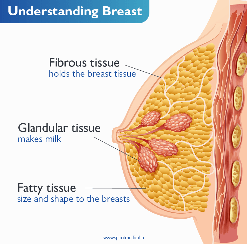
There are three parts in a woman’s breast:
Fibrous tissue: This holds the breast tissue in place.
Glandular tissue: This part makes milk and is called the lobes. Ducts are the tubes responsible for carrying milk to the nipple.
Fatty tissue: This is responsible for giving size and shape to the breasts. This tissue fills up the space between the fibrous tissue, lobes and ducts.
The breast can be dense or non-dense. Dense breast means fat is less and fibro-glandular tissue is more. In non-dense breasts, fat is more.
What is a mammogram?
According to CDC, a mammogram refers to an X-ray picture of the breast to confirm early signs of breast cancer.
How mammogram is done?
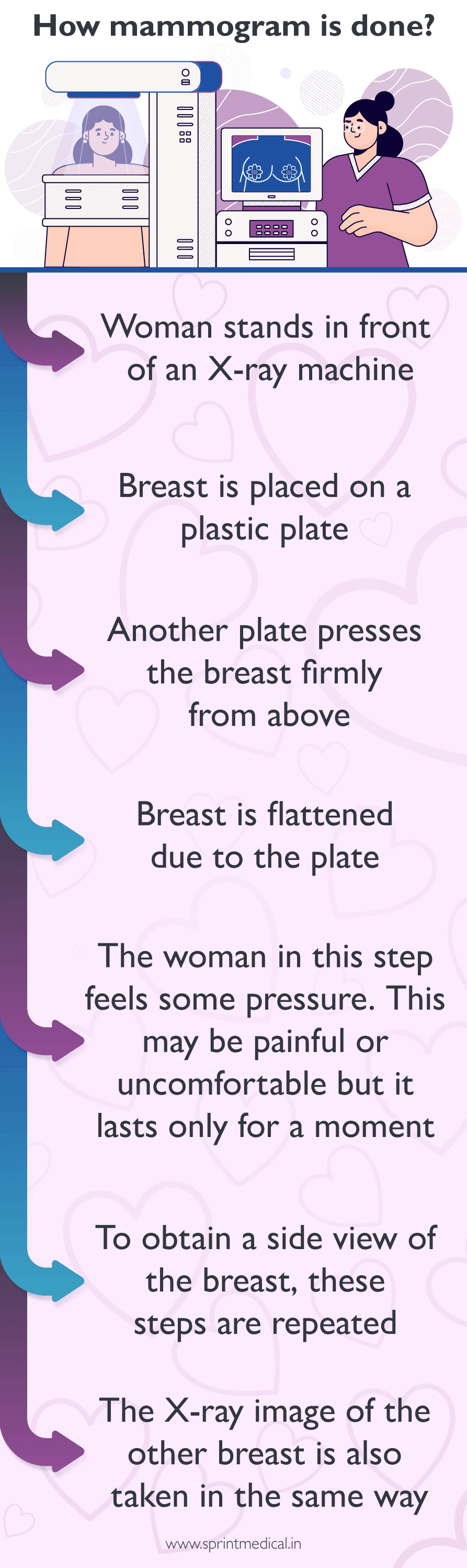
The procedure is given below:
Woman stands in front of an X-ray machine, which is specialised.
Breast is placed on a plastic plate.
Another plate presses the breast firmly from above.
Breast is flattened due to the plate.
The woman in this step feels some pressure. This may be painful or uncomfortable but it lasts only for a moment.
To obtain a side view of the breast, these steps are repeated.
The X-ray image of the other breast is also taken in the same way.
Important considerations in effective conversation while planning mammograms
Every woman must be aware of some important aspects of her breast health. Some must-know points about women’s health are given below:
Every woman must be aware of her breast health.
An open and frank discussion with family and friends is always important. This increases awareness and inspires you more towards your health.
Note down the questions you want to ask when you schedule an appointment with your doctor.
Do not believe in myths and do not follow them while planning conversation.
Women need to know their first menstruation date.
For post-menopausal women, it is important to remember the last menstruation date and record of hormonal treatment if taken.
We must know our weight.
Always discuss your medical history with the doctor.
Preparation before mammogram:
Proper preparation is required if you plan to get your first mammogram done. Let us understand it thoroughly with reasons.
Mammograms should be scheduled a few days after the menstrual cycle.
Mammograms are not scheduled a week before the periods because the breast may be tender or swollen.
Deodorants, lotion, cream and powder in the breast and underarm areas must not be used on the day of mammograms. This is because these are shown up as white spots on the X-ray.
The woman should wear loose clothes and feel relaxed. Dress should be avoided because the woman needs to undress from the waist up for the procedure.
Do not wear jewellery as it distorts the mammogram images.
Clear all doubts about mammograms:
It is essential to know the points below.
Mammogram is safe.
It involves low-dose radiation.
The woman may feel uncomfortable for a brief duration.
Mammograms are painless for most women.
Planning after a normal mammogram:
After getting mammogram results as normal, it is important to ask the following questions from your doctor.
Ask the doctor when to have the next mammogram.
Take your opinion if you have any questions related to diet, type of exercises, etc.
Planning after an abnormal mammogram:
If you and your loved one get an abnormal mammogram, it may concern you, but it is important to know and understand the points below:
Do not panic because every lump is not cancerous.
Wait for more test results.
Doctor may advise additional mammogram, ultrasound, MRI and biopsy to confirm malignancy.
Breast density in mammography report:
It is important to know breast density.
It reflects the amount of fibrous and glandular tissue present in the breasts as compared to the fatty tissue.
Breast density is seen on a mammogram. Dense breasts are seen in nearly half of the women aged 40 years or older.
The categories of breast density as given on the CDC website are:
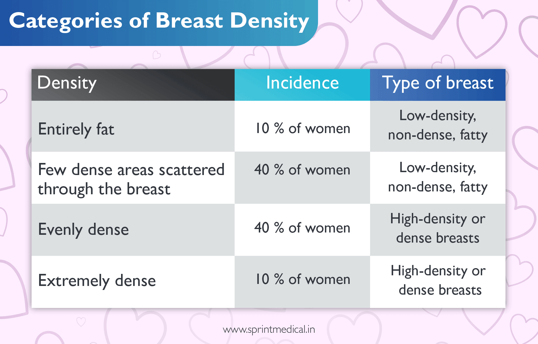
One important question every woman must ask after a mammogram: Are my breast dense?
A dense breast appears white on a mammogram, while a non-dense breast appears dark. When a woman has dense breasts, the chances of breast cancer are higher, and the diagnosis may be masked on the mammogram when the white area abnormally appears more. Cancer is detected as white on a mammogram.
Women are likely to have dense breasts at a young age, during pregnancy, breastfeeding and with low body weight. A woman undergoing hormone replacement therapy also has dense breasts.
According to National Cancer Institute, women aged 40 and older may experience dense breasts on mammograms. So, every woman after 40 must pledge for a yearly mammogram.
Breasts change over time. This change is hormonal. This is what happens after menopause; the breast tissue changes, and after the age of 55 chances of breast cancer detection are more.
There are some other causes of dense breasts, like family history, weight loss, and hormonal therapy. It is always important to talk about breast health.
Dense breast and 2D mammogram: Is diagnosis complete?
Women with dense breasts must not rely only on 2D mammograms for diagnosis.
Another gold standard exists to aid in detecting and confirming breast cancer diagnosis. They are
3D mammograms
Ultrasound: In breast ultrasound, sound waves are used, and pictures are obtained of areas inside the breast. These are called sonograms.
MRI: Breast MRI (Magnetic Resource Imaging) provides detailed pictures of areas inside the breast. This is a type of scan.
Digital breast tomosynthesis (DBT):
FDA approved the first Digital Breast Tomosynthesis (DBT) unit on February 11, 2011.
This is the fourth generation of mammography technology
DBT, also known as 3D mammography, is an advanced mammogram technology which is specifically useful in detecting breast cancer in dense breast tissue.
Standard mammography involves 2D breast images, whereas 3D mammography provides three-dimensional images of the breast.
The U.S. Food and Drug Administration (FDA) approved digital breast tomosynthesis (DBT) in 2011
The diagnosis of breast cancer in the dense breast is challenging. There are chances that malignancy may get missed with conventional 2D mammography, but it could be easily detected in 3D mammography.
DBT takes multiple breast pictures from more angles in an arc-like pattern, thereby reducing the frequency of a false-positive result.
How is DBT done?
The breast is placed and compressed the same way as done in the standard mammogram procedure.
In DBT, the arm of the X-ray tube moves in an arc over your breast. As it moves, it takes many 2D images from multiple angles, digitised and combined to obtain a 3D image.
The image is more detailed compared to a standard mammogram.
Steps in Digital Breast Tomosynthesis:
Stand in front of the mammography machine.
One of the breasts is placed on the platform.
Plastic plate squeezes the breast against the platform.
You feel pressure and discomfort.
Stay still when the machine is moving.
Hold your breath for a few seconds.
X-ray tube moves in an arc-like motion.
Several images of breasts are obtained.
Reconstruction of images into multiple slices of 1mm thickness.
images are viewed on a monitor approved for tomosynthesis.
Procedure is repeated for another breast.
Take-Home Points
This article clears all the doubts you may have while planning for your first mammogram.
A mammogram is a completely safe procedure and is a standard diagnostic procedure in breast cancer screening. However, 3D mammograms or Digital Breast tomosynthesis are more sensitive to diagnosing breast cancer.
You may feel discomfort, but compression helps to obtain a clearer view of your breast tissue. It also allows for a lower dose of radiation.
Breast health is an important aspect of women’s health so it is important to know the points discussed in the article so that you become more aware and take the right step towards the right direction.
FAQ on Mammogram
At the age of 40, it is recommended to get your first mammogram done.
It may detect cancer early before the appearance of symptoms. It provides high-quality images.
In a 2D mammogram, one image is taken from the top and the other from the side, while in a 3D mammogram, multiple images are taken, which provide a more detailed picture of the breast.
Yes, Mammography, including tomosynthesis, involves the use of the lowest and safest radiation dose.
Tomosynthesis is more sensitive in detecting cancer in dense breast tissue.
The duration is 2-3 seconds for a standard mammogram.
The duration is 4 seconds.
Tomosynthesis.
Both techniques use low-dose X-rays.
Comments ( 0 )
No Comments
Leave a Comment
Related Posts

Food requirement for lactating Mother | Diet for Breastfeeding Mother
Breast milk is filled with protective compounds and nourishing nutrients essential for the healthy development of your baby. As a mother, your requirements for nutrients increase for nutrient-dense and nourishing foods to aid your breast milk production.


Mazia Ahmed

Snack Recipes for Breastfeeding Moms!
“Bottles fill his stomach, but breastfeeding fills his soul” To keep his soul happy moms need to be fit and healthy, so here are some quick one-handed snacks for Breastfeeding Moms

Juveriya Anwar Momin
Health & Wellness Tips
Subscribe to our blog